
Fig. 14.
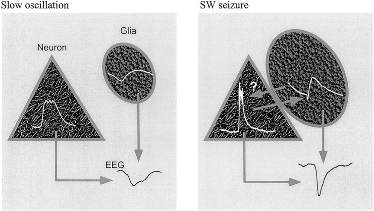
Schematic diagram of the mechanisms generating field potentials during slow oscillations (left) and SW seizures (right). An averaged cycle is drawn in each cell (white traces). During the slow oscillation, reversed neuronal and glial potentials contribute to the genesis of the extracellular field potential (EEG). SW seizures are accompanied by glial swelling, which may bring patches of cellular membranes into contact, allowing intraneuronal potentials to appear reversed, as field potentials, in the glial cells (arrowfrom neuron to glia points toward the glial negativity). The reverse pathway might also be at work (arrow from glia to neuron). Both intraneuronal and intraglial activities contribute to the shape of the extracellular field potential.