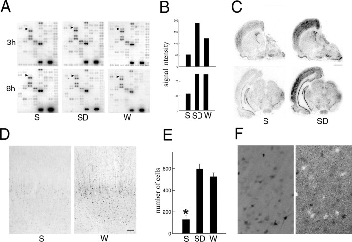
Fig. 2.
Arc expression in the cerebral cortex after sleep and waking. A, cDNA microarrays showing cortical Arc mRNA levels (arrowheads) after 3 hr (3h) or 8 hr (8h) of sleep (S; n = 7), sleep deprivation (SD; n = 7), and waking (W; n = 6).B, Densitometric analysis performed by scanning the microarrays with a PhosphorImager. The y-axis values refer to signal intensity (arbitrary units). C,In situ hybridization for Arc mRNA in brain sections of a representative rat killed after 8 hr ofS and of a rat killed at the same circadian time after 8 hr of SD. Scale bar, 1.5 mm. D, Arc levels measured with immunocytochemistry in the parietal cortex of rats after 3 hr of S and 3 hr of W. Scale bar, 100 μm. E, Mean number (±SEM) of Arc-immunoreactive neurons in parietal cortex after 3 hr ofS (n = 8), SD(n = 8), and W(n = 5). The sampled area was a 500-μm-wide cortical column spanning all layers (Mann–Whitney Utest, *p < 0.01). F, Double labeling in the parietal cortex of a rat that was sleep deprived for 3 hr. Arc immunoreactivity (black cells;left) does not colocalize with parvalbumin immunoreactivity (white cells; right). Scale bar, 50 μm.

