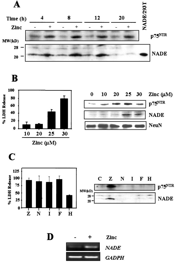
Fig. 1.
Co-induction of p75NTR and NADE in cultured cortical neurons after zinc exposure. A, Western blots for p75NTR and NADE (22 kDa). Samples were prepared from cortical cultures at indicated hour after 15 min exposure to 300 μm zinc. Positive control for NADE was prepared from 293T cells transfected with NADE (NADE/293T) (Mukai et al., 2000). B, Left, Bars represent LDH release (mean + SEM; n = 3) in cortical cultures, after 24 hr exposure to indicated concentrations of zinc.Right, Western blots for p75NTR and NADE (22 kDa) in cortical cultures after 12 hr exposure to indicated concentrations of zinc. Western blots for the neuronal marker NeuN were used as control. C, Left, Bars represent LDH release (mean + SEM; n = 4–8), 16 hr after 15 min exposure to 300 μm zinc (Z) or continuous 16 hr exposure to 30 μm NMDA (N), 500 nm ionomycin (I), 100 μmFeCl2 (F), or 50 μmH2O2 (H). All but H2O2 induced near complete neuronal death.Right, Western blots for p75NTR and NADE 4 hr after 15 min exposure to zinc or after 4 hr exposure to other toxicants as above. Whereas zinc (300 μm, Z) induced both, NMDA, ionomycin, FeCl2, or H2O2 induced neither. D, RT-PCR assays for NADE mRNA in cortical cultures, sham-washed, or 8 hr after 15 min exposure to 300 μm zinc. RT-PCR assay for GAPDH mRNA is presented as control.

