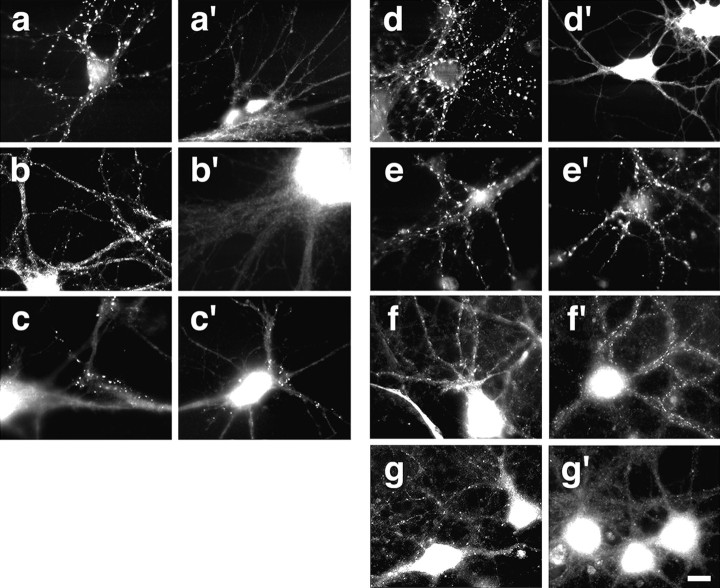
Fig. 3.
Effects of the antisense oligonucleotide treatment on wild-type and agrin-deficient neurons. Immunocytochemical analysis of synaptic differentiation in 10-d-old, antisense oligonucleotide-treated hippocampal neurons. a–c′, Rat (P3–P5) hippocampal neurons were treated with 7.5 μm AS oligonucleotides. Patterns of immunoreactivity are shown for synapsin-I (a, a′), synGAP (b, b′), and the NMDA receptor subunit NR1 (c, c′). The treatment with the AS oligonucleotides affected the synapsin-I and synGAP staining but not the NR1 immunoreactivity. d–e′, E18 mouse hippocampal neurons were treated for 10 d with 7.5 μm AS oligonucleotides. Synapsin-I immunoreactivity is shown in untreated (d, e) and treated (d′, e′) neurons. Synapsin-I-immunoreactive puncta disappeared in AS-treated wild-type (d′) but not in AS-treated agrin-deficient (e′) neurons. f–g′, synGAP staining in wild-type (f, f′) and agrin-deficient (g, g′) E18 hippocampal neurons is shown. Cultures were maintained for 13 d (f, g) and 24 d (f′, g′). Although synGAP puncta disappeared in mutant neurons between 13 and 24 d, they were still observed in wild-type neurons. The pattern of synapsin-I immunoreactivity did not change during this time period (data not shown). Scale bar, 20 μm.

