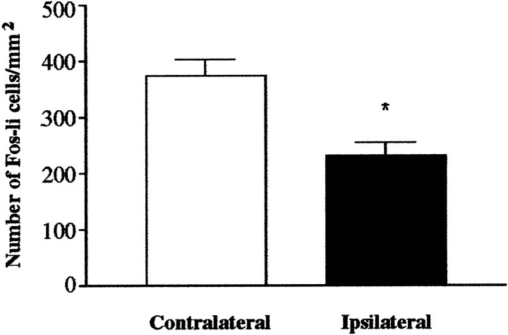
Fig. 8.
NMDA lesions of the thalamus decrease the number of Fos-li neurons in the cortex in response to DOI challenge. The number of Fos-li neurons in the SSC on the side of the lesion drops sharply relative to the intact side. There was no significant difference (p = 0.200) between the number of Fos-li neurons in the contralateral (intact) SSC of sham- and lesioned-treated rats. *p 163 ≤ 0.001 relative to intact (contralateral) side.

