Fig. 1.

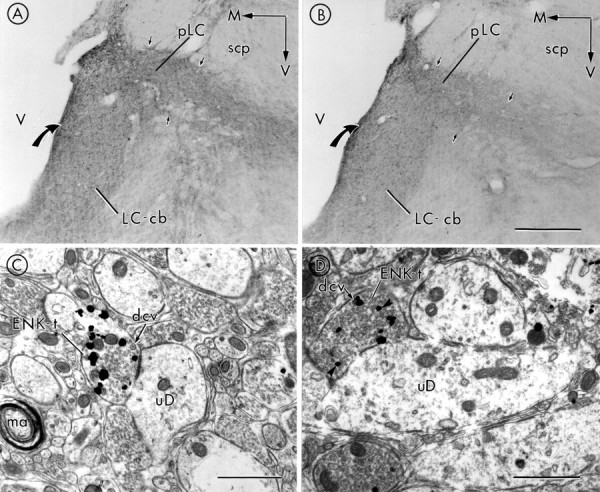
Photomicrographs showing peroxidase labeling for ENK in the caudal aspect of the LC cell body region (cb) (also defined as nuclear or core of the LC) and peri-LC (pLC) areas (region containing noradrenergic dendrites of LC neurons) of placebo- and morphine-treated rats.A, Peroxidase labeling for ENK can be detected in varicose processes in LC and peri-LC areas in a placebo-treated rat. Varicose processes are distributed throughout the region of the LC containing noradrenergic cell bodies (LC-cb) as well as in peri-LC areas (small black arrows), which are known to contain noradrenergic dendrites of LC neurons.B, There is a decrease in peroxidase immunoreactivity for ENK in the LC-cb region as well as in the peri-LC ventral to the superior cerebellar peduncle (scp;small black arrows) in morphine-treated animals. For both Aand B, a large black curved arrow points to the medial aspect of the LC immediately adjacent to the ventricle (V). Arrows point medially (M) and ventrally (V). C, D, Electron micrographs showing immunogold–silver labeling for ENK in axon terminals in the LC (ENK-t) of a placebo-treated rat (C) and a morphine-treated rat (D). Note that in C there are more gold–silver particles indicative of ENK immunolabeling in an axon terminal apposed to an unlabeled dendrite (uD), as compared with the ENK-t in the opiate-dependent rat (D). ENK-t in both C andD contains numerous small, clear vesicles as well as large, dense-core vesicles (dcv). Scale bars:A, B, 250 μm; C, 0.94 μm; D, 0.75 μm. ma, Myelinated axon.
