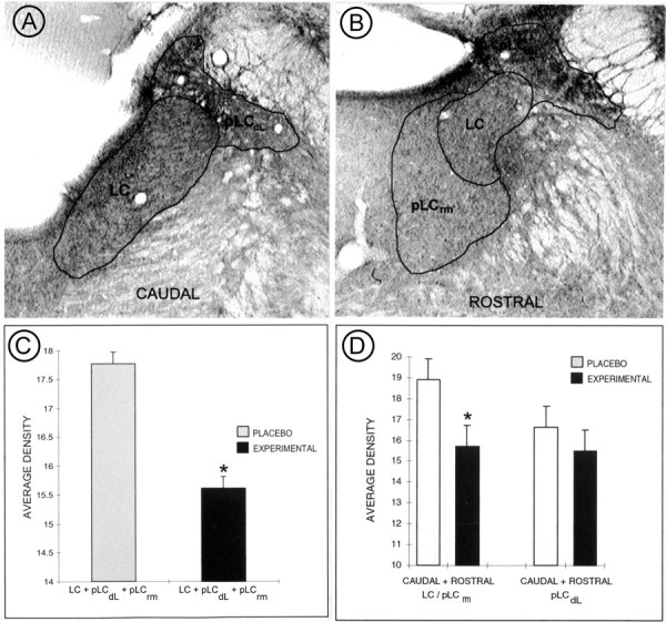
Fig. 2.

Light level densitometry measurements of coronal tissue sections taken from placebo- and morphine-treated rats that were processed for immunoperoxidase localization of ENK. A,B, Representative sampling of regions defined as LC (which comprises the core of the LC) and peri-LC (which comprises the dorsolateral aspect, pLCdL, as well as the rostromedial, pLCrm) at both caudal and rostral levels of the dorsal pons of control rats. The nuclear LC consists of portions of the dorsal pontine tegmentum containing noradrenergic cell bodies, whereas the peri-LC areas contain noradrenergic dendrites. Because the ENK labeling is extensive in peri-LC areas and extends into the medial parabrachial, sampling was restricted to portions of the neuropil known to contain noradrenergic dendrites. C,D, Bar graphs illustrating quantification of pixel values obtained from LC and peri-LC areas shown in A andB. In C, the values for the LC and pLC were combined, whereas in D the samples are shown for caudal + rostral LC/pLCrm and caudal + rostral pLCdL areas. Note that there is a statistically significant decrease in peroxidase immunolabeling in samples obtained from morphine-treated rats. There is also a statistically significant decrease in the caudal + rostral LC/pLCrm obtained from morphine-treated rats.
