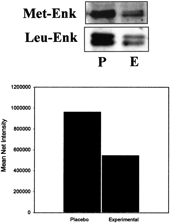
Fig. 4.
Western blot analysis of samples obtained from the LC of placebo-treated (P) and experimental (E) morphine-treated rats. Expression of the primary antibody directed against met- or leu-ENK was readily detected in microsamples obtained through the LC region. Multiple bands were detected on the gel; however, proteins that migrated above the 33.9 kDa marker most likely represent the migration of proenkephalin-like opioid peptides. Expression was reduced in samples of the LC obtained from morphine-dependent rats (E). The bar graph represents the mean net intensity observed from Western blots (n = 6) processed for met-ENK from placebo- and morphine-treated rats.

