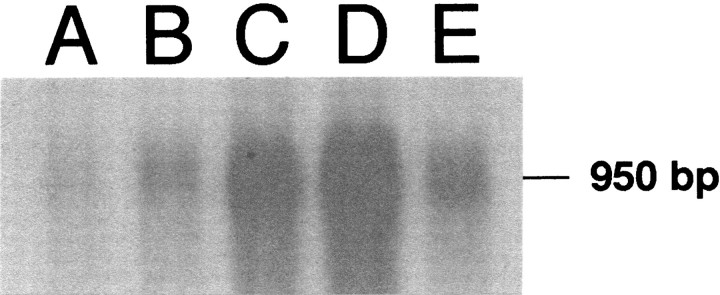
Fig. 5.
Northern blot showing a 950 bp band indicative of PPE mRNA in three different tissue specimens: medulla oblongata containing the nucleus paragigantocellularis (PGi) from morphine-treated (lane A) and placebo-treated (lane B) rats, striatum from morphine-treated (lane C) and placebo-treated (lane D) rats, and heart (lane E, used as a positive control for PPE mRNA). Plasmids were successfully transformed into anEscherichia coli strain, and the insert DNA was isolated from the vector by restriction digestion using SacI andSmaI enzymes. Note that the level of PPE mRNA is reduced in lane A, representing medullary regions containing the PGi of morphine-treated rats as compared with a similar sample obtained from placebo-treated rats (lane B).

