
Fig. 1.
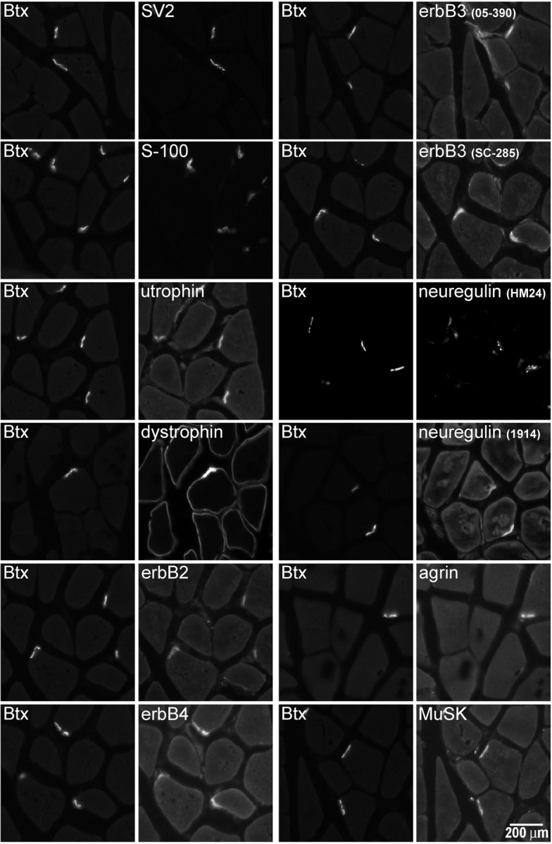
Immunofluorescence visualization of proteins enriched at the NMJ. Proteins of the agrin/MuSK and neuregulin/erbB signaling pathways as well as marker proteins for subdomains of the NMJ were visualized in cross sections of adult rat gastrocnemius muscle using a 20× objective. Bodipy-αBtx was used to identify individual NMJs. The axon terminal marker SV2, Schwann cell marker S-100, utrophin (a protein enriched in the postsynaptic primary gutter), and dystrophin (a protein enriched in the secondary folds) all show clear staining at the NMJ. Dystrophin additionally shows the expected enrichment along the sarcolemma. Immunofluorescence from antibodies against erbB2, erbB3, erbB4, neuregulin, agrin, and MuSK all show enrichment at the NMJ with little extra-junctional staining of the muscle. Scale bar, 200 μm.