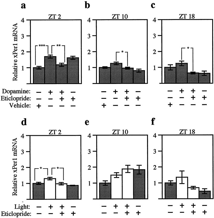
Fig. 6.
Three hour pulses of dopamine and light have modest effects on xPer1. Northern blots from the experiments described in Figure 5 were hybridized with anxPer1 probe. Three hours of dopamine (a–c; 500 nm) or light (d–f; 3–5 × 10⁻⁴ W/cm2) were delivered to eyecups beginning at early subjective day (a, d; ZT 2), late subjective day (b, e; ZT 10), or subjective night (c, f; ZT 18) after previous treatment with 50 μm eticlopride, a D2-like dopamine antagonist. The abundance of xPer1 mRNA was quantitated as in Figure2c, and values were normalized to the vehicle-alone group for each experiment. The mean value of each group was calculated from four to five retinas from individual animals. Error bars indicate SEM. White bars indicate those groups receiving light;graybars indicate those groups maintained in constant darkness. At ZT 2 (a), the dopamine-treated group was significantly different from the vehicle-alone group (***p < 0.001, ANOVA). At all three times of day (a–c), the dopamine-treated groups were significantly different from the dopamine plus eticlopride groups (**p < 0.01, *p < 0.05, ANOVA). At ZT 2 (d), the light-treated group was significantly different from both the control group and the light plus eticlopride group (*p < 0.05, ANOVA). No significant differences were detected between the light-treated groups and either the control groups or the light plus eticlopride groups at ZT 10 or ZT 18 (e, f; ANOVA).

