
Fig. 3.
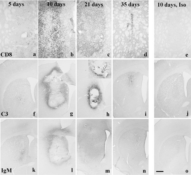
a–e, CD8 staining in xenografts and isografts. a, Five-day-old xenografts contain a few CD8+ cells and by (b) 10 d the staining is intense and extends outside the borders of the graft. c, It is still present at 21 d after implantation, although it wanes as the graft is lost such that (d) by 35 d only a few CD8+ cells are seen scattered around the site of the original graft. e, In contrast, a 10-d-old isograft has no CD8+ cells within it. f–j, Complement (C3) staining in isografts and xenografts. f, There is minimal complement staining in the xenograft at 5 d after implantation. In contrast (g) there is clear staining in and around the graft at 10 d that (h) persists at day 21 and is more clearly seen at higher power (inset). i, By day 35 the staining has become less intense as the graft is rejected. j, In contrast, 10-d-old isografts have no such complement binding.k–o, Rat-specific IgM staining in isografts and xenografts. k, There is some IgM staining in the xenograft at 5 d after implantation. l, By 10 d it has become more apparent and is around and within the graft.m, At 21 d the staining is diffuse and involves the whole hemisphere, (n) a pattern of staining that is also seen at 35 d. o, In contrast, a 10-d-old isograft induces no such IgM binding. Scale bar (shown ino): a–e, 0.5 mm; f–o, 1 mm.