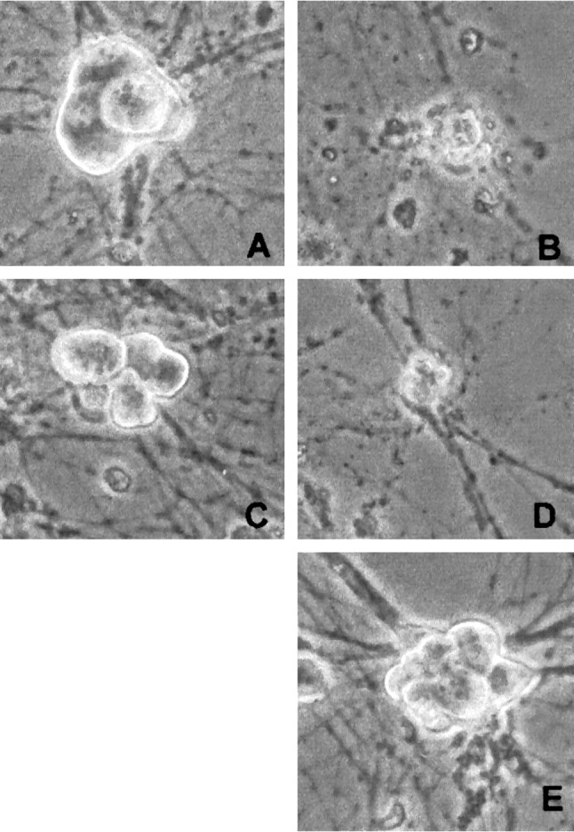
Fig. 10.

Phase-contrast micrographs of rat sympathetic neurons maintained in NGF-containing medium and exposed to and/or infected with the following: no treatment (A), UV (B), (Δ1–126) DN DP1 virus + UV (C), (Δ1–126) DN DP1 control virus + UV (D), or (Δ1–126) DN DP1 virus alone (E). The photos were taken 2 d after irradiation and/or infection.
