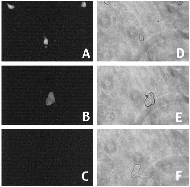
Fig. 11.
Immunofluorescence (A–C) staining with an antibody directed against the FLAG epitope or corresponding light micrographs (D–F) of sympathetic neurons in culture infected with Sindbis virus expressing (Δ233–272) DP1F (A, D) or (Δ103–126) DN DP1F (B, E) or containing a nonexpressing control virus (C, F). Neurons were stained 2 d after infection.

