
Fig. 4.
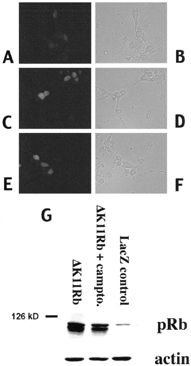
Immunofluorescence (A–F) and Western immunoblot (G) analyses of ΔK11 Rb expression. A—F, Immunofluorescence (A, C, E) staining with an antibody directed against pRb or corresponding light micrographs (B, D, F) of cortical neurons in culture infected with ΔK11 Rb-expressing virus (C–F) or control (A, B). Neurons were fixed and stained with anti-pRb antibody 24 hr after infection (A–D) and after 12 hr of camptothecin treatment (E, F). G, Western immunoblot analysis of whole-cell extracts of cortical neurons infected with adenovirus expressing ΔK11 Rb (no treatment or 10 hr camptothecin treatment) or LacZ as indicated. The blots were analyzed using an anti-pRb antibody, stripped, and reprobed with anti-β-actin.