
Fig. 6.
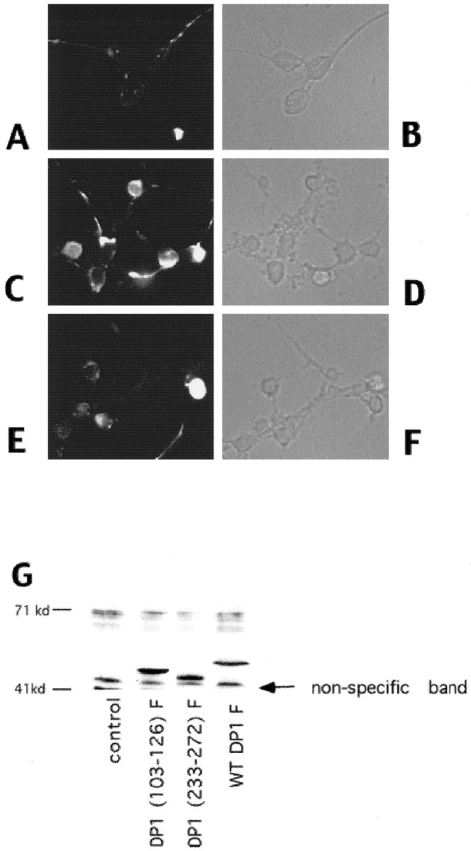
A—F, Immunofluorescence (A, C, E) or corresponding phase (B, D, F) images of neurons infected with control virus (A, B) or with virus encoding (Δ103–126) DN DP1F (C–F). Neurons were fixed and stained with anti-FLAG antibody 24 hr after infection (A–D) and after 12 hr of camptothecin treatment (E, F). G, Western immunoblot analyses of whole-cell extracts of cortical neurons infected with Sindbis viruses expressing WT or the indicated mutant FLAG-tagged DP1 constructs. control indicates samples in which the neurons were infected with a control virus. Theblots were analyzed using an anti-FLAG antibody. A nonspecific immunoreactive band is marked as indicated. Neurons were lysed 24 hr after infection with the indicated viruses.