Fig. 1.
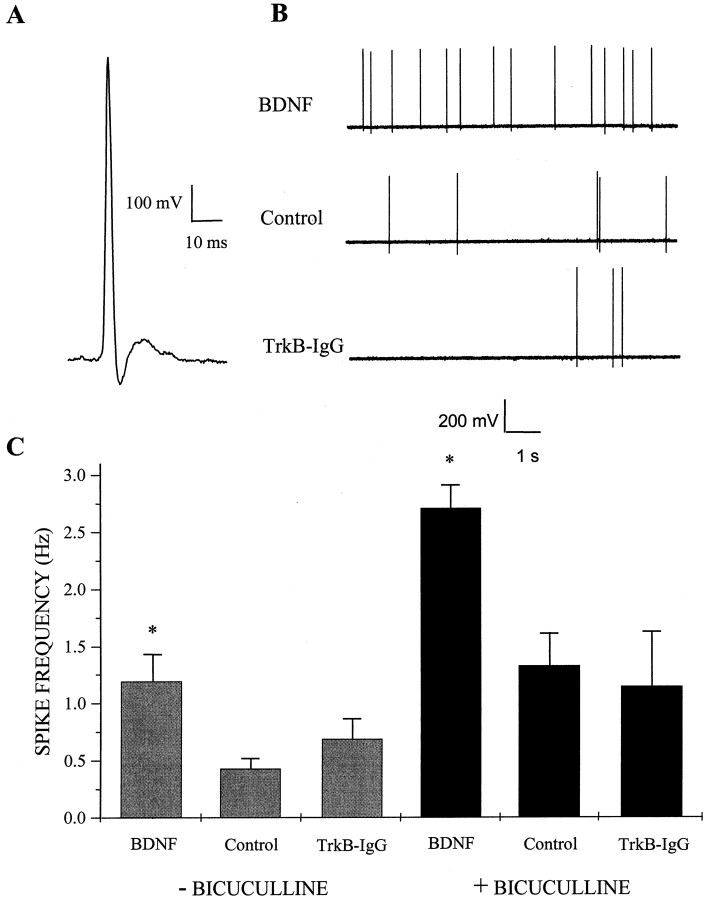
BDNF increases spontaneous action potential firing rate. A, Representative traceillustrating the waveform of an action potential recorded with an on-cell patch pipette; this configuration was used to measure action potential frequency in dissociated hippocampal cultures. Voltage traces were inverted about the vertical axis to conform to convention. Data were acquired at 5 kHz in voltage-follower recording mode and filtered at 2 kHz. B, Spontaneous action potential firing was increased in cultures treated with BDNF. Representative on-cell recordings shown at a compressed time base from cells treated for 4–7 d with 100 ng/ml BDNF (top), untreated (Control, middle), or treated with 5 μg/ml TrkB-IgG (bottom). Cultures were rinsed in recording saline several times to ensure that no BDNF or TrkB-IgG was present at the time of recording. C,Left, BDNF treatment increased the spontaneous firing rate of pyramidal neurons approximately threefold compared with untreated controls; means ± SEM are shown. *p< 0.009 indicates a significant difference by ANOVA between BDNF and either control or TrkB-IgG treatment groups;n = 33, 27, and 36 for BDNF, control, and TrkB-IgG groups, respectively. Right, Elevated action potential firing rates induced by BDNF persisted in disinhibited circuits. BDNF appeared to increase excitatory synaptic transmission directly because the increase in spontaneous firing rates of pyramidal neurons persisted after acute blockade of inhibitory transmission by bicuculline during the recording period. *p < 3 × 10⁻⁵ indicates a significant difference by ANOVA between BDNF and either control or TrkB-IgG treatment groups;n = 43, 36, and 12 for BDNF, control, and TrkB-IgG groups, respectively.

