
Fig. 1.
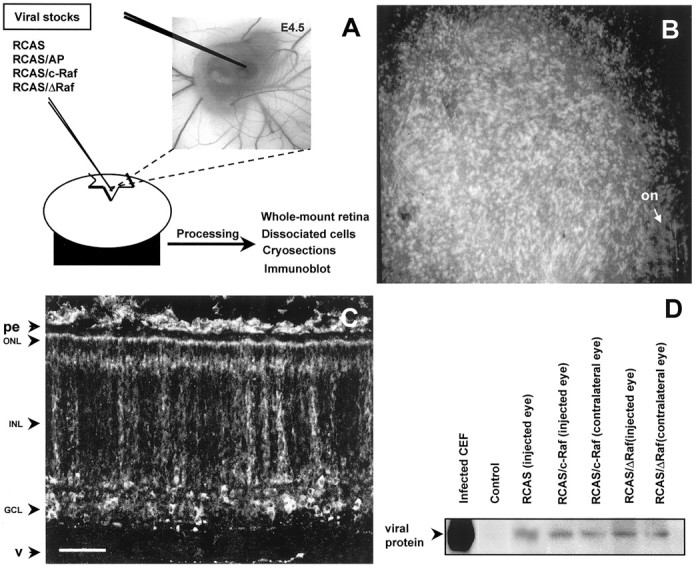
Retroviral infection of chick embryonic retina.A, Schematic representation of the viral injection at E4.5, as described in Materials and Methods. Viral infection was monitored by immunostaining with the monoclonal antibody anti-Gag19, as shown in B–D. B, A low-magnification micrograph of a whole-mount stained infected retina. C, A stained retinal cryosection shows the widespread, clonal-reminiscent distribution of the infected cells. The retinal layers are indicated:on, optic nerve head; pe, pigmented epithelium; ONL, outer nuclear layer;INL, inner nuclear layer; GCL, ganglion cell layer; v, vitreous humor. D, A representative immunoblot of infected neuroretinas. Compare the expression levels between the injected and the contralateral eye of the same embryo. Infected chick embryonic fibroblasts (CEF) and the retina of a noninjected embryo (Control) are shown. Scale bar: B, 1 mm; C, 50 μm.