
Fig. 4.
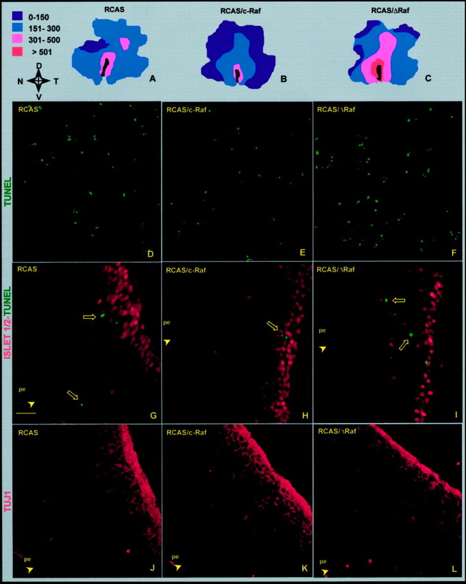
Effect of the interference with Raf on apoptosis and the morphogenesis of the ganglion cells. Retinas injected at E4.5 with the indicated viral constructs were processed for TUNEL in whole-mount retina (A–F) or for immunostaining in retinal cryosection (G–L) 48 hr after injection. Retinas with total dead cell scores closest to the average value (see Fig. 3) were represented as isothanas (A–C; the pseudocolor scale indicates dead cell density per square millimeter). The orientation of the retinas is indicated:N, nasal; T, temporal; D, dorsal; V, ventral. Comparative fields in the temporoventral quadrant were obtained by confocal microscopy of the represented retinas (D–F). Double-stained cryosections for the neuronal cell marker Islet-1/2 (red) and apoptotic cells by TUNEL (green) (G–I). Note that, in the control infection with empty vector (G), at this age, Islet-1/2 is a selective nuclear marker of ganglion cells located in their proper layer. Serial sections stained for the ganglion cell marker TUJ1, which also stains the optic fiber layer (J–L). In all cases, only sections including the lens and the optic nerve were chosen, and temporal fields 0.5 mm away from the optic nerve head are shown. The pigmented epithelium (pe) side is indicated. Scale bar:A–C, 1.5 mm; D–F, 40 μm;G–L, 20 μm.