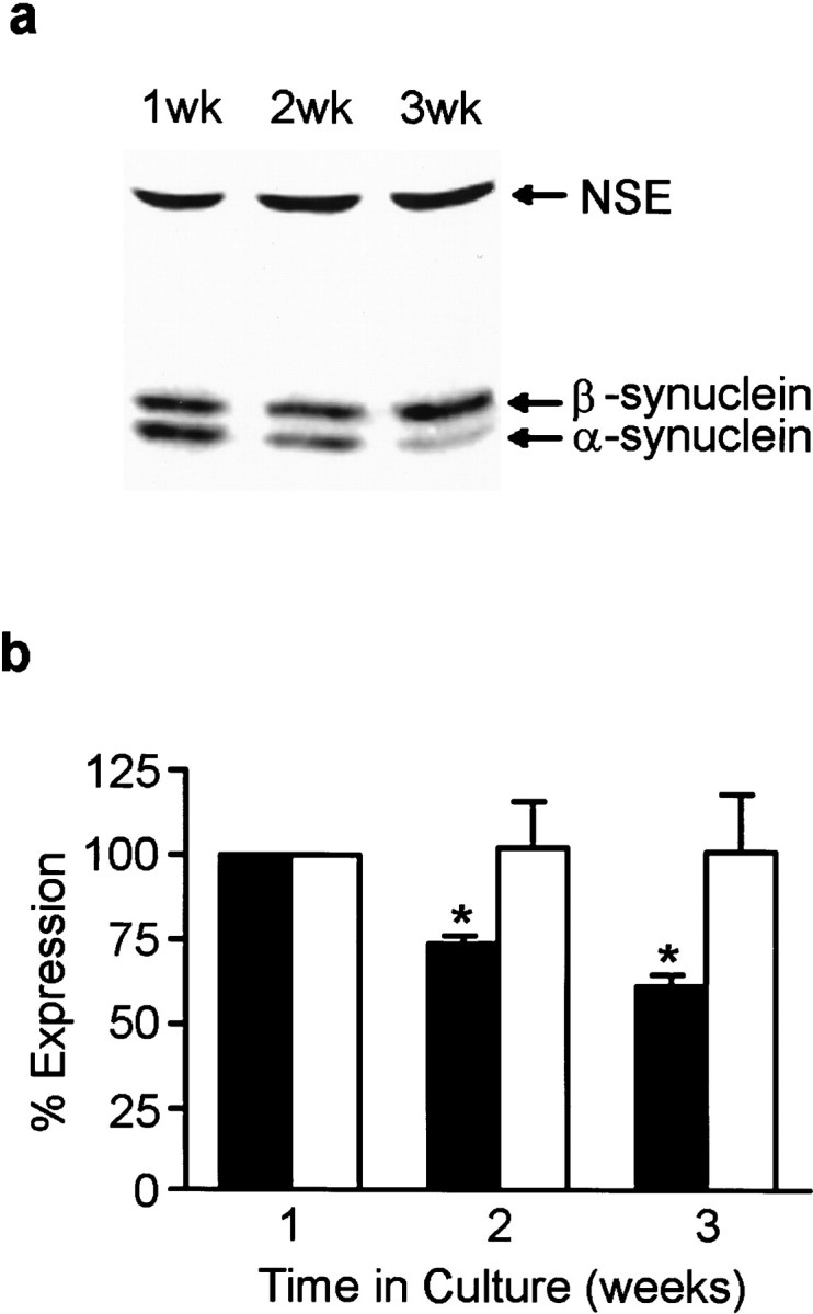
Fig. 2.

Synuclein protein expression in cultured hippocampal neurons. α- and β-synuclein expression in neurons at 1, 2, 3 weeks in culture was determined by quantitative Western blot analysis from Triton X-100 soluble cell extracts (a). The blots were probed with antibody syn102 (specific for both α- and β-synucleins) and an antibody to NSE.b, Quantitation of α- and β-synuclein expression at 1, 2, and 3 weeks in culture. Closed bars represent α-synuclein expression, and open bars represent β-synuclein expression. All synuclein signals were normalized to the signal for NSE to account for the amount of neuronal protein loaded per lane. Results are presented as the average percentage of expression ± SEM. The expression at 1 week was arbitrarily set at 100%. n = 3; *p < 0.05.
