
Fig. 2.
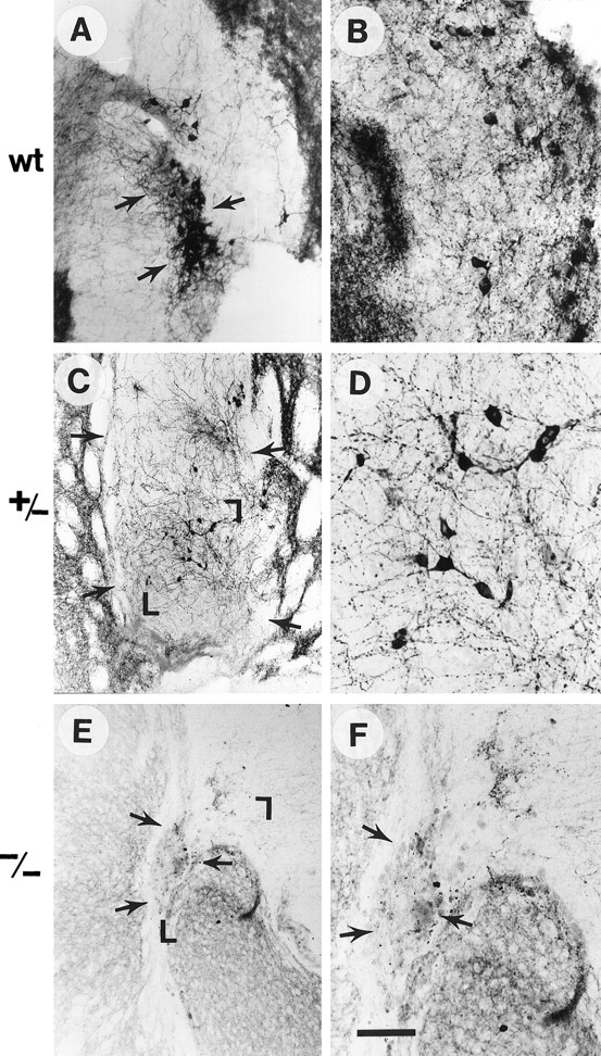
TH immunocytochemistry of grafts from WT (A, B), GDNF +/− (C, D), and GDNF −/− (E, F) donors. D andF are larger magnifications of the grafts shown inC and E, respectively, whereasB represents a high magnification of a wild-type graft other than the one seen in A. Arrowsdelineate grafts, and the magnified areas are demarcated with corners in C and E. Note the much greater TH-immunoreactive cell numbers and fiber outgrowth in WT compared with −/− grafts, which were virtually devoid of TH-positive neurons and neurites and, additionally, contained a large number of macrophages. The +/− grafts had an intermediate number of TH-positive cells and fibers. Scale bar (shown in F): A, 70 μm; C, E, 100 μm;B, D, F, 30 μm.