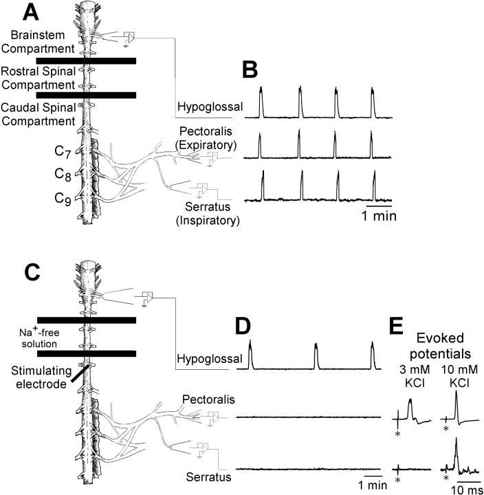
Fig. 1.
Spontaneous respiratory motor activity and electrically evoked potentials produced by turtle brainstem-spinal cord preparations. A, C, Drawings of turtle brainstem–spinal cord preparations are shown with barriers between C2 and C3 and between C4 and C5, which establish the brainstem compartment, rostral spinal compartment, and caudal spinal compartment.B, Suction electrodes attached to hypoglossal nerve rootlets and intact pectoralis and serratus nerves record rhythmic, synchronized spontaneous respiratory motor activity. The integrated traces (time constant, 200 msec) are shown for each nerve.C, Schematic drawing of the turtle brainstem–spinal cord preparation with Na+-free solution flowing into the rostral spinal cord compartment, resulting in a blockade of action potential conduction between the brainstem and spinal cord.D, Under these conditions, suction electrodes attached to hypoglossal nerve rootlets record rhythmic, spontaneous respiratory activity, whereas electrodes attached to pectoralis and serratus nerves record no respiratory activity. E, A metal electrode inserted into the spinal cord at C5 ipsilateral to the intact pectoralis and serratus nerves (dark heavy linein C) was used to electrically stimulate descending synaptic inputs to pectoralis and serratus spinal motoneurons (stimulus artifacts marked with asterisks). Evoked potentials were typically observed in pectoralis, but not serratus, nerves when the caudal spinal compartment [K+] was 3 mm (left traces). When the caudal spinal compartment [K+] was 10 mm(right traces), pectoralis evoked potentials were larger (stimulus artifacts in both pectoralis traces are the same absolute size; the gain on the left trace is 5 times larger than that on the right trace), and evoked potentials appeared on serratus.

