
Fig. 2.
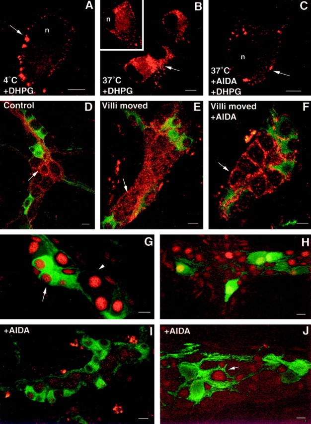
Agonist- and reflex-evoked internalization of mGluR5 in enteric neurons. A–C, Confocal images showing the localization of mGluR5 immunoreactivity (arrows) in dissociated neurons. Neurons were incubated with DHPG (30 μm) for 1 hr at 4°C, washed, and incubated at 37°C for 0 min (control; A), 30 min (B), or in the presence of AIDA (C). Arrows in Bindicate internalization of mGluR5 from the plasma membrane to the cytoplasm. D–F, Confocal images showing the localization of mGluR5 in submucosal neurons in tissue incubated without agitation (D), in response to villous agitation (E), and in response to villous movement in the presence of AIDA (F). Internalized receptor (red) is found in the cytoplasm of submucosal neurons that do not (arrowhead) contain ChAT immunoreactivity (green). G–J, pCREB immunoreactivity in enteric neurons in response to distension in the absence (G, H) or presence (I, J) of AIDA. G, pCREB-immunoreactive nuclei (red) are found in ChAT-positive (green; arrow) and ChAT-negative (arrowhead) submucosal neurons (green). H, A group of calbindin-immunoreactive neurons (green) displays pCREB immunoreactivity (red) in the myenteric plexus.I, J, AIDA blocks the expression of pCREB (red) in ChAT-immunoreactive submucosal neurons (I; green) and in calbindin-immunoreactive myenteric neurons (J;green); however, a subset of calbindin-negative myenteric neurons continue to express pCREB (J;arrow). Scale bars, 30 μm.