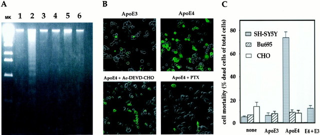
Fig. 2.
Characterization of ApoE4 toxicity.A, DNA laddering. F11 cells were treated with 30 μg/ml of ApoE4 (lane 2) or 30 μg/ml of ApoE3 (lane 3) for 24 hr. Cells were also treated with 30 μg/ml of ApoE4 in the presence of 10 μm Ac-DEVD-CHO (lane 4), 1 μg/ml PTX (lane 5), or 30 μg/ml of ApoE3 (lane 6) for 24 hr. Extracted DNA was applied onto a 2% agarose gel (2 μg/lane). As another control, DNA extracted from F11 cells grown in a complete growth medium was examined for DNA ladder formation (lane 1). MK indicates a molecular marker, which is a StyI digestion product of λ (cI857Sam7) DNA. This marker gives a 500–600 bp interval in the bottom half of a gel. The results shown are representative of three similar experiments independently performed.B, TUNEL assay. F11 cells were treated with 30 μg/ml of ApoE3 or ApoE4 for 24 hr; cells were stained with TUNEL. In the bottom panels, cells were treated with 30 μg/ml of ApoE4 in the presence of 1 μg/ml PTX or 10 μmAc-DEVD-CHO for 24 hr. Phase-contrast images were superimposed on the TUNEL fluorescence images. The fragmented DNA was stainedgreen by TdT and TUNEL. Apoptotic cells are known to undergo leakage of DNA from nuclei, which allowed cellular staining of DNA as well as nuclear staining. The results shown are representative of three similar experiments independently performed.C, Effect of ApoE in other cell lines. SH-SY5Y cells, Bu695 glial cells, or CHO cells were treated with or without 30 μg/ml of ApoE3 or ApoE4, and cell mortality was measured by Trypan blue exclusion assay. In SH-SY5Y cells, 30 μg/ml of ApoE4 was also treated in the presence of 30 μg/ml ApoE3 (the extreme right lane). The incubation periods were 48 hr for SH-SY5Y and CHO cells and 72 hr for Bu695 cells. CHO cell mortality at 72 hr after 30 μg/ml ApoE4 treatment was similar to that at 48 hr after treatment (data not shown).

