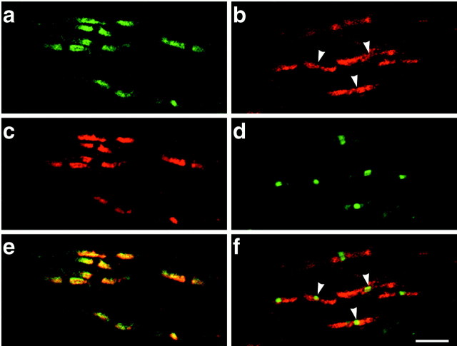
Fig. 2.
Contactin is localized in the paranodes and nodes in the optic nerve. Staining of contactin with βC–Fc (a) and an anti-Caspr antibody (c) demonstrated specific colocalization of these proteins at the paranodes (merged in e). Staining with an affinity-purified anti-contactin antibody (b) demonstrates that contactin in the paranodes frequently extends through the nodes of Ranvier; an antibody to ankyrinG (d) identifies the nodes in the same field. In the merged images (f), contactin that is colocalized with ankyrinG at the node appears yellow; nodes in which contactin is readily visible are indicated by the white arrowheads. Scale bar, 5 μm.

