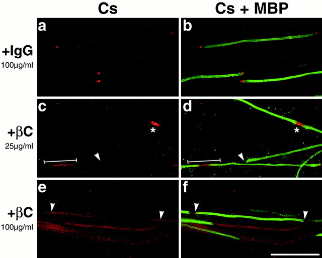
Fig. 8.
βC–Fc perturbs Caspr distribution in myelinating cocultures. Cocultures were maintained in myelinating media supplemented with human IgG (a, b) as a control or with media supplemented with either 25 μg/ml (c, d) or 100 μg/ml (e, f) of βC–Fc and stained for Caspr and MBP. Control cultures (100 μg/ml IgG) displayed the normal paranodal distribution of Caspr (Cs; rhodamine) and normal myelin formation (MBP; fluorescein). βC–Fc-treated cultures (+βC) myelinate normally but exhibit concentration-dependent abnormalities of Caspr localization, including the absence of Caspr at the paranodes (white arrowheads), Caspr staining at the node (asterisk), and Caspr staining throughout the internode (white line). At the highest concentration, Caspr was absent from essentially all paranodes. Scale bar, 50 μm.

