
Fig. 1.
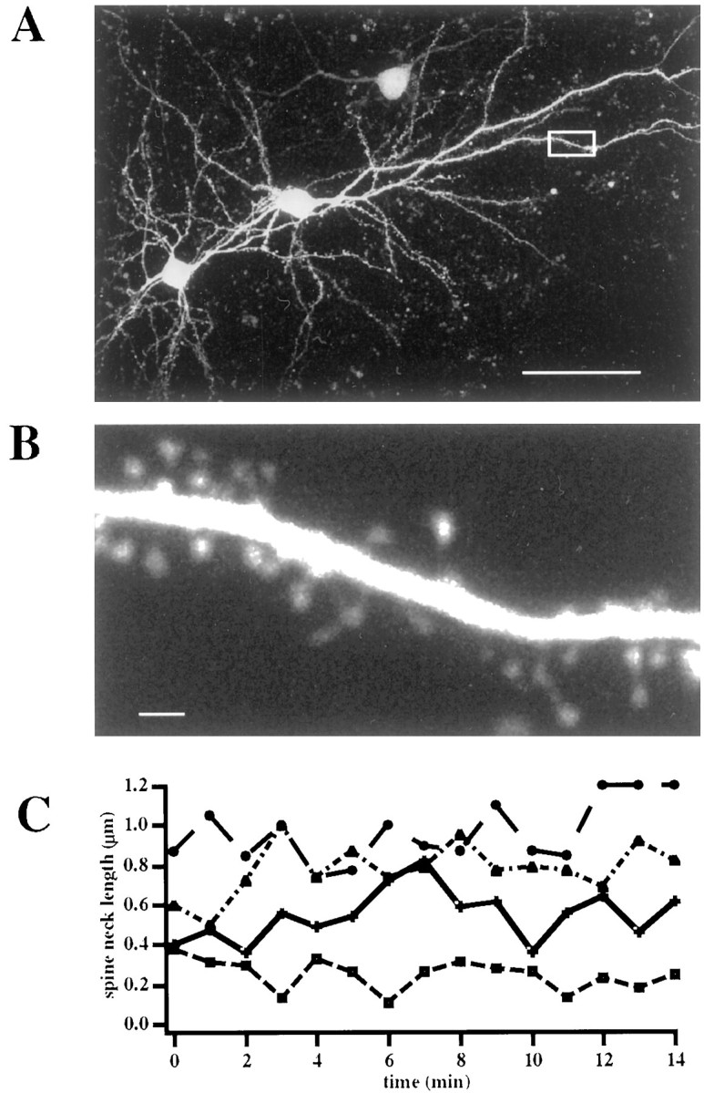
Spine neck lengths change during spine movement.A, Two-photon image of two EGFP-transfected layer 2/3 pyramidal neurons. The pial surface is to the top right.B, High-magnification image of the boxed area in A. Note how dendritic spines are clearly visible. C. Spine necks elongate and retract as they move. Analysis of the spine neck changes in four spines followed for a 15 min period. Scale bars: A, 50 μm; B, 1 μm.