Fig. 4.
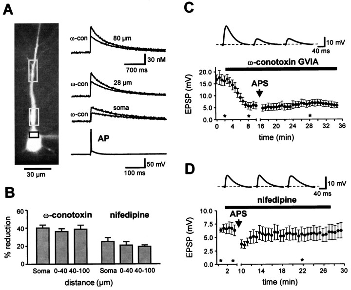
Associative LTD is dependent on Ca2+ influx via both, voltage-gated N- and L-type Ca2+ channels. A, Fluorescence image of a CA1 pyramidal neuron filled with 0.1 mm fura-2 (380 nm excitation). Rectangles indicate somatic and dendritic ROIs from where Ca2+ transients were recorded with high time resolution (100 Hz, traces on the right). The action potential-induced Ca2+ transients were reduced in the presence of 1 μm ω-conotoxin GVIA (ω-con, N-type blocker). B, The bar graph summarizes the mean inhibition of the Ca2+ transient by ω-conotoxin GVIA (n = 6) obtained with 0.1 mm fura-2 and 10 μm nifedipine (n = 5; L-type blocker) obtained with 0.1 mm Oregon Green (see Materials and Methods). Dendritic ROIs were located in the stratum radiatum at a distance of ∼0–40 μm or 40–100 μm from the soma. C, Application of 0.5 μm ω-conotoxin GVIA (n = 9) reduced basal synaptic transmission and prevented the induction of LTD by APS.D, A 10 μm concentration of nifedipine did not affect basal transmission but similarly inhibited LTD induction (n = 7). Voltage traces in C andD are EPSPs from a single representative experiment, respectively, at the times indicated by theasterisks.

