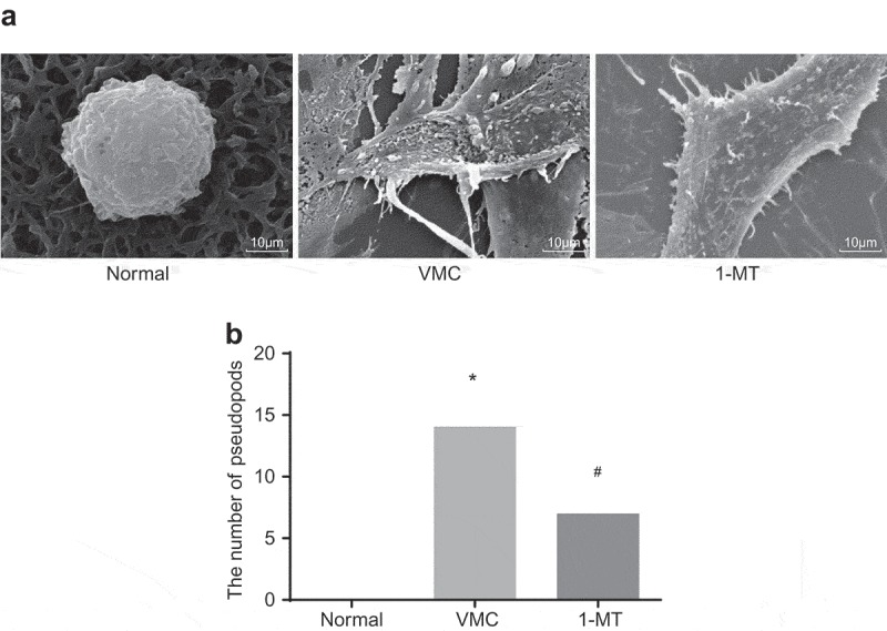
Figure 4.

1-MT treatment impairs the activation of macrophages in VMC mice (× 8000). A, images of macrophages in abdominal cavity in normal, VMC and 1-MT treated VMC mice observed by SEM; B, the quantitative analysis of the number of pseudopods in normal, VMC and 1-MT treated VMC mice; * p< 0.05 vs. normal mice without any treatment; # p< 0.05 vs. CVB3-induced VMC mice. Statistical values were measurement data and analyzed using one-way ANOVA. n = 20 in the VMC group. n = 40 in the normal group. n = 14 in the 1-MT group. 1-MT was the inhibitor of IDO1. The experiment was repeated 3 times independently. VMC, viral myocarditis; SEM, scanning electron microscopy; CVB3, coxsackievirus B3; ANOVA, analysis of variance.
