Figure 1.

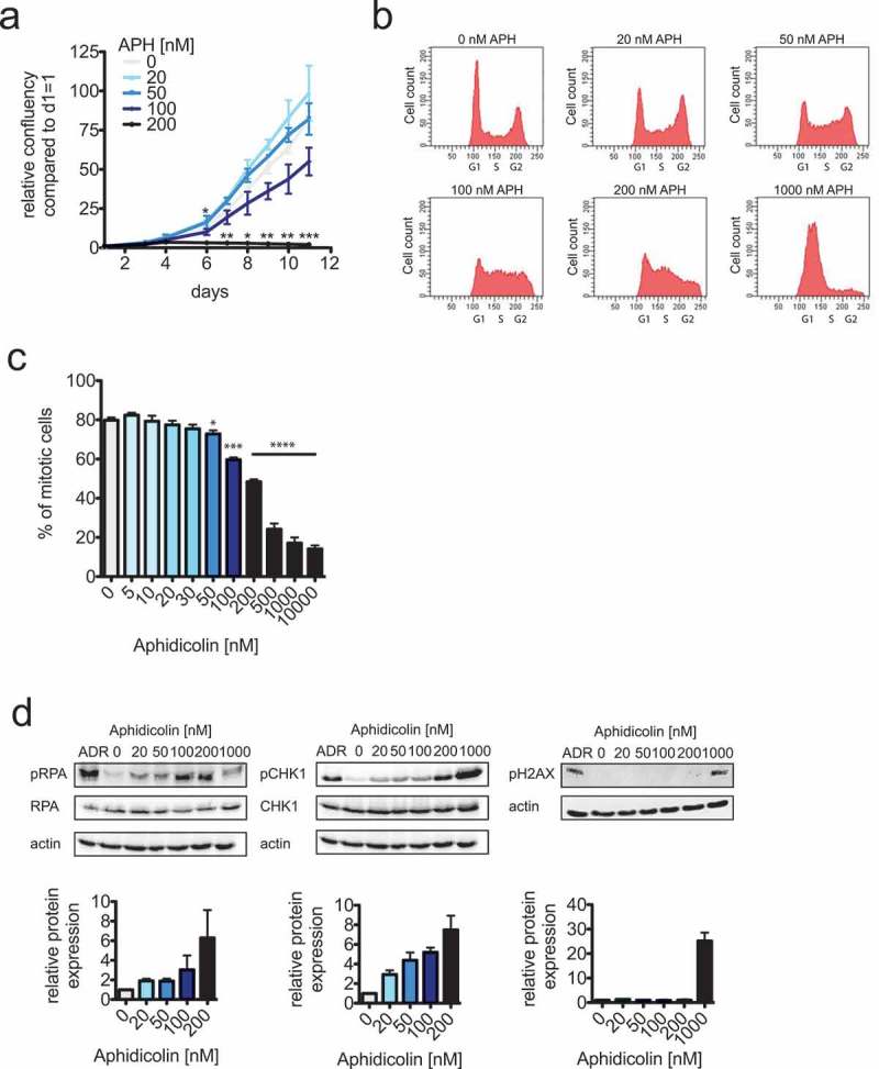
Only very mild replication stress allows efficient cell proliferation.
(a) Cell proliferation measurements of HCT116 cells treated with increasing concentrations of aphidicolin (APH) for up to 11 days. Cells were seeded at identical cell numbers, treated with increasing concentrations of aphidicolin and cell proliferation was quantified using a Celigo cytometer based on area confluency (n = 3 experiments, mean ± SEM, t-test). (b) Representative FACS profiles of HCT116 cells treated with increasing concentrations of aphidicolin (APH) for 24 hours. Cells were stained with propidiumiodide and the DNA content was determined.(c) Determination of mitotic entry of HCT116 cells treated with increasing concentrations of aphidicolin. Asynchronously growing cells were pre-treated with aphidicolin for 4 hours and subsequently together with dimethylenastrone for additional 20 hour to arrest cell cycle progression beyond prometaphase. Mitotic cells were quantified by FACS analyses detecting phosphorylated MPM2 epitopes (n = 3 experiments, mean ± SEM, t-test). (d) Western blot analyses to detect phospho-S33-RPA, phospho-S345-Chk1 and phospho-Ser139-H2AX as markers for S/G2 checkpoint activation and DNA damage. HCT116 cells were treated with the indicated aphidicolin concentrations or with 600 nM adriamycin (ADR) to induce DNA damage for 24 hours. Whole cell lysates were subjected to western blot detecting the indicated antigens. Representative western blots are shown and band intensities were quantified based on three independent experiments.
