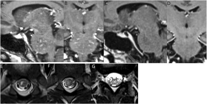
Figure. Sagittal (A) and coronal (B) 3D T1-weighted postcontrast images showing punctate and curvilinear enhancement in the pons.
Follow-up sagittal (C) and coronal (D) 3D T1-weighted postcontrast images approximately 3 weeks later showing resolution of enhancement after treatment with IV methylprednisolone and rituximab. Axial T2-weighted images of the thoracic spinal cord (E and F) and conus (G) at the time of presentation showing patchy T2 hyperintense lesions involving gray and white matter.

