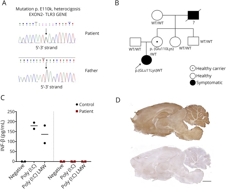
Figure. Genetic, functional, and immunologic studies in a 6-year-old child with TLR3-pathway deficiency who developed HSE, and a relapse of the viral infection followed by AE post-HSE.
(A and B) Genetic studies identified a missense heterozygous mutation p.Glu110Lys (c.328G>A) in exon 2 of the TLR3 gene (NM_003265.2) in the patient and her mother (who was asymptomatic). The patient's grandfather had history of severe recurrent herpetic keratitis but at the time our patient was studied, he was deceased, and no genetic studies were available. (C) IFN-β production by fibroblasts of the patient (red squares) and a healthy control (black dots) in 2 independent experiments after 24 hours stimulation with 2 different TLR3 agonists: Poly(I:C) or Poly(I:C) LMW; stimulation with its own culture media was used as negative control (negative). IL-6 production by TLR3-agonists stimulated MDDCs was also decreased in the patient compared with a healthy control in 2 independent experiments (data not shown). (D) Rat brain immunostaining with CSF of the patient (upper panel) indicating the presence of antibodies against neuronal surface antigens, compared with that of a healthy control individual (lower panel). Scale bar = 2,000 μm. AE = autoimmune encephalitis; HSE = herpes simplex encephalitis; IFN-β = interferon-beta; LMW = low molecular weight; MDDC = monocyte-derived dendritic cell; Poly(I:C) = polyinosinic-polycytidylic acid; TLR3 = Toll-like receptor 3.

