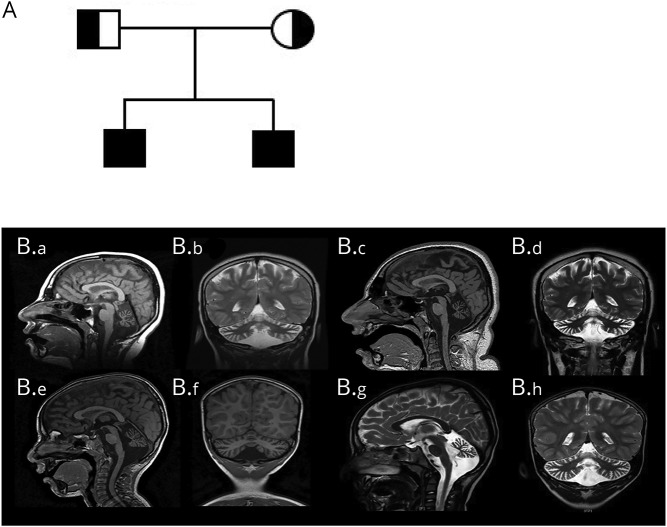
Figure 1. Pedigree and brain imaging.
(A) Family pedigree. (B) Neuroimaging findings in the affected siblings. (B.a–B.d) Serial brain imaging for sibling 1. (B.a) Sagittal T1 image at 9 years showing normal cerebrum and brainstem, with diffuse cerebellar atrophy and moderately dilated fourth ventricle with prominent CSF spaces surrounding the cerebellum. (B.b) Coronal T2 image at 9 years showing cerebellar atrophy of both hemispheres and vermis. (B.c) Sagittal T1 and (B.d) coronal T2 images for sibling 1 at the age of 16 years, shows similar findings of stable severe cerebellar atrophy. (Be–B.h) Serial brain imaging for sibling 2. (B.e) Sagittal T1 image at 3 years showing cerebellar volume loss with prominent CSF spaces and fourth ventricle; brain and brainstem are normal. (B.f) Coronal T1 at 3 years showing small vermis and cerebellar hemispheres. (B.g) Sagittal T2 and (B.h) coronal T2 images for sibling 2 at the age of 5 years showing stable cerebellar atrophy.

