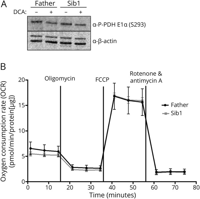
Figure 4. Analysis of PDH E1α phospho293 (S293) expression and oxygen consumption rates in parent and BRAT1 patient primary fibroblasts.

(A) Representative Western blot analysis showing no observed differences in PDH E1α S293 expression in parent (Father) and BRAT1 patient (Sib1). Protein lysates were extracted from untreated cells or fibroblasts treated with 5 mM DCA (B) OCRs of the same fibroblast cells using the Seahorse XFe96 Extracellular Flux Analyzer (Seahorse Bioscience). OCR was normalized to protein to eliminate differences in growth rate between cell lines (n = 8 wells/cell line). DCA = dichloroacetate; PDH = pyruvate dehydrogenase; OCR = oxygen consumption rates.
