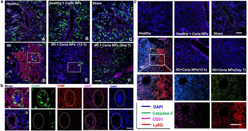
Figure 4. Immunofluorescence staining on liver samples.
a Immunofluorescence staining was performed using anti-CLE4F antibody (green) as Kupffer cell marker, anti-F4/80 antibody (red) as monocyte/macrophage marker, anti-CD31 antibody (pink) as an endothelial marker, and DAPI (blue) for nuclear staining of liver tissues from each group. Scale bar: 100 µm. b Enlarged images of immunofluorescence staining in a from liver samples of IRI mice treated with PBS (i) or ceria NPs (ii). Scale bar: 50µm. Yellow oval dash line marked the cross-section of blood vessel in liver. c. Neutrophil marker was stained by using anti-Ly6G antibody (red), while using anti-caspase-3 antibody (green) as a cell apoptosis marker, anti-CD31 antibody (pink) as an endothelial marker, and DAPI (blue) for nuclear staining of liver tissue from each group. Scale bar: 100 µm.

