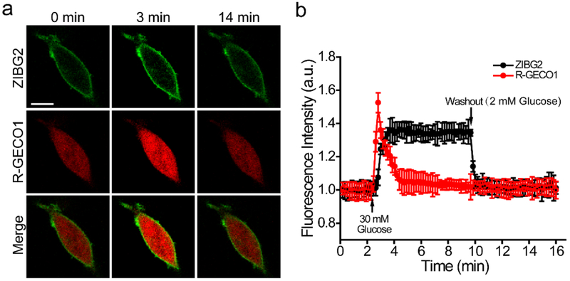
Figure 5.
Fluorescence imaging of glucose-induced Zn2+ and Ca2+ dynamics in MIN6 cells. (a) Fluorescence images of cells expressing ZIBG2 on the cell surface and R-GECO1 intracellularly. Scale bar: 20 μm. (b) Quantitative results for intensity changes of ZIBG2 and R-GECO1 presented as mean and s.d. of 8 cells from three independent replicates. The arrow indicates the time point for addition of high glucose (30 mM) and washout with low glucose (2 mM).

