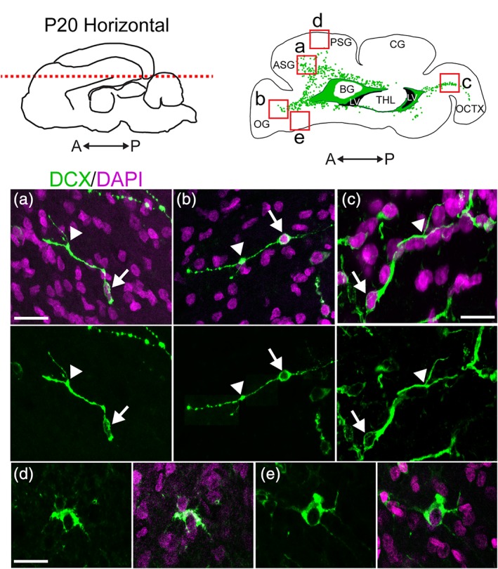
Figure 2.

DCX+ cells in the P20 ferret cortex and white matter exhibit different morphologies. (a–c) DCX+ cells in the white matter exhibit a migratory morphology including an elongated cell body (arrow) and a single, sometimes forked, leading process (arrowhead). (d,e) DCX+ cells in the cortex have large, rounded cell bodies, large nuclei, and multiple extended processes. Scale bar a, b, d, e = 10 μm. Scale bar c = 7.5 μm [Color figure can be viewed at http://wileyonlinelibrary.com]
