Figure 5.

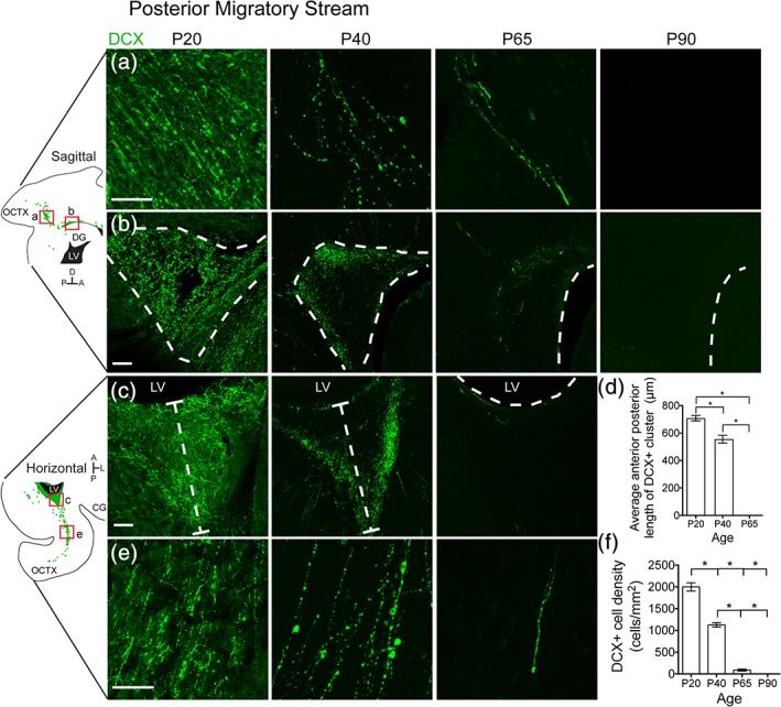
The ferret posterior migratory stream disappears between P40 and P65. (a,b) P20 sagittal section highlighting the PMS. (a) Individual DCX+ cells with migratory morphology oriented away from the lateral ventricle and toward the occipital cortex. (b) DCX+ cluster posterior to the lateral ventricle. (c,e) P20 horizontal section highlighting the PMS. (c) DCX+ cluster posterior to the lateral ventricle. (d) Quantification of the decrease in DCX+ cluster size over time. (e) Individual DCX+ cells with migratory morphology oriented away from the lateral ventricle and toward the occipital cortex. (f) Quantification of the decrease in DCX+ cell density over time. Scale bar a–c,e = 100 μm [Color figure can be viewed at http://wileyonlinelibrary.com]
