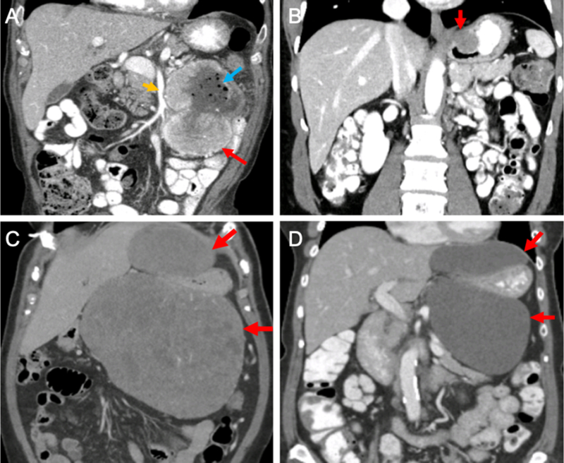
Figure 1 -. Examples of primary GISTs.

(A) Coronal CT image of a proximal jejunal GIST (red arrow) near the superior mesenteric artery (orange arrow). Note the air bubbles within the tumor (blue arrow), indicating the existence of a sinus tract between the bowel and the tumor. (B) Coronal CT image of a GIST (red arrow) located at the gastroesophageal junction. (C&D) Coronal CT images of a massive gastric GIST (C) before and (D) after 6 months of neoadjuvant imatinib. Red arrows indicate the bilobed tumor surrounding the stomach. Note the decreased density of the tumor after therapy. At operation, the tumor arose from the gastric cardia and was removed with splenectomy and a limited partial gastrectomy. CT – computed tomography
