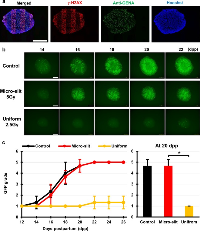
Figure 3.
Tissue-sparing effect of micro-slit radiation in spermatogenesis. (a) Immunohistochemical images of ex vivo testes culture tissues 1 h after 10 Gy 200 μm-slit X-ray microbeam irradiation. Staining for γ-H2AX, Anti-GENA (anti-germ cell-specific antigen antibody), and Hoechst (for DNA) is shown as red, green, and blue, respectively. Scale bars, 500 μm. (b) Representative images show Acr-GFP expression changes in single cultures following 0 Gy (control), 5 Gy micro-slit (50%), and 2.5 Gy uniform (100%) X-ray irradiation, from 14 to 22 days postpartum (dpp). Scale bars, 500 μm. (c) Chronological changes in Acr-GFP expression after the micro-slit and the uniform X-ray irradiation. A minimum of three tissue samples each from different donor mice were used for each experiment. Data represent the mean GFP expression ± SD. Asterisk indicates P < 0.01.

