Figure 3.
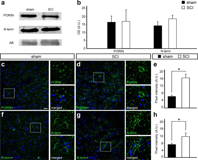
Western blot and immunofluorescence analysis of voltage-dependent anion-selective channel 1 (VDAC1) 24 hours after spinal cord injury (SCI) counterstained with 4′-6-diamino-2-phenylindole (DAPI). (a) Representative western blot bands using anti-VDAC1 PORIN, anti-VDAC1 N-term in the spinal cord samples of sham controls and SCI animals. Amido black bands were used as loading controls. (b) Quantification of optical densities from the bands obtained using anti-PORIN and anti-N-term in spinal cord samples from sham controls and SCI animals. Representative image of the ventral region of the spinal cord from (c) sham controls and (d) SCI animals stained with anti-PORIN (green) and counterstained with DAPI (blue), with high magnification view of the respective selected area. (e) Graph indicating the pixel intensity of anti-PORIN labeling in sham controls and SCI animals. Representative image of the ventral region of the spinal cord from (f) sham controls and (g) SCI animals stained with anti-N-term (green) and counterstained with DAPI (blue), with high magnification view of the respective selected area. (h) Graph indicating the pixel intensity of N-term labeling in sham controls and SCI animals. Bars represent standard error of the mean. Scale bar: 50 μm. *P < 0.01.

