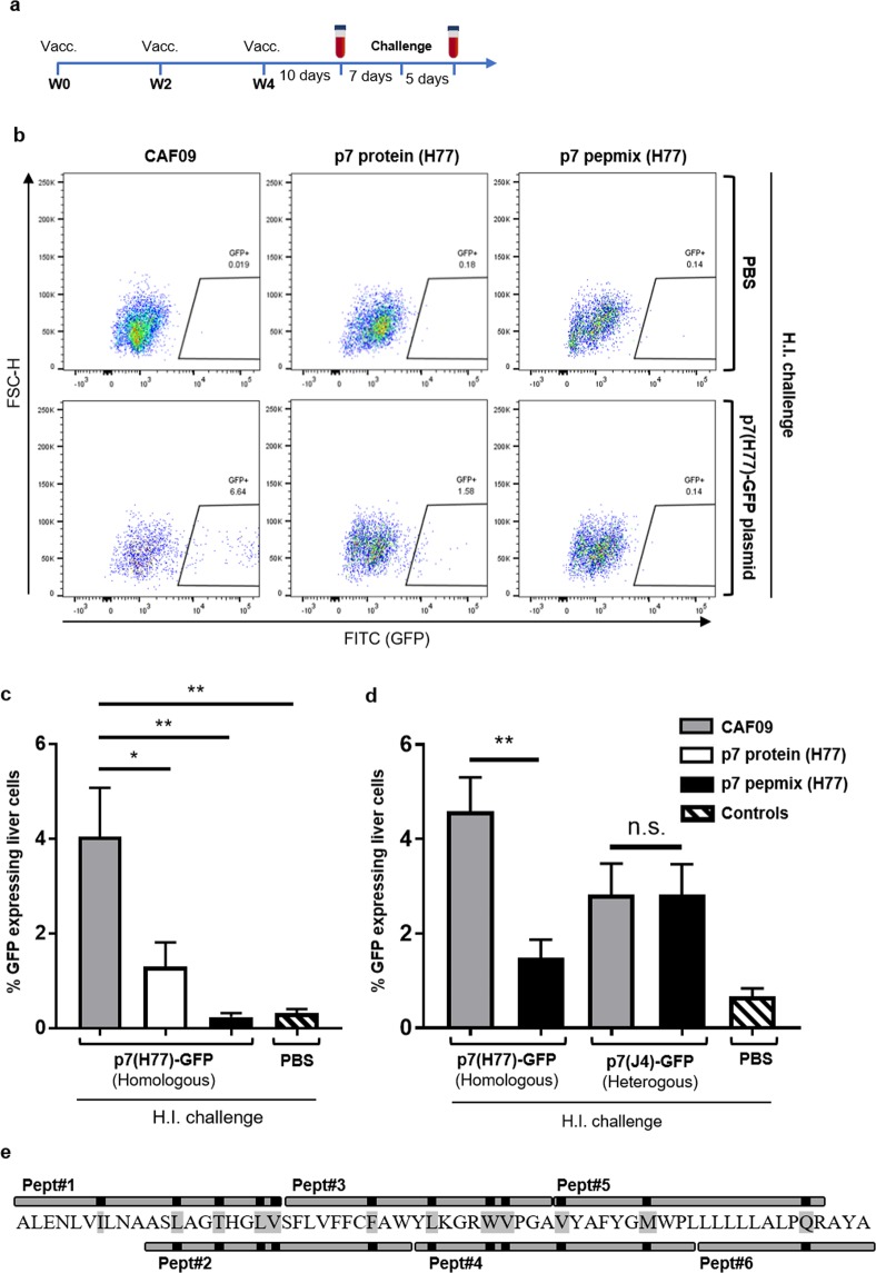
Figure 3.
HCV p7 vaccination reduced GFP-expression in the liver after surrogate challenge. (a) The diagram indicates time points for vaccination, bleeding, surrogate challenge and termination of the experiment. (b) Representative FACS-plots shows gating of GFP-expressing liver cells from CAF09 versus p7 protein and pepmix (strain H77) vaccinated mice five days after hydrodynamic injection (H.I.) of PBS (upper panel) or 100 µg p7(H77)-GFP plasmid (lower panel). (c) The bar charts show frequencies of GFP-expressing liver-cells in vaccinated mice after surrogate challenge. Control mice that received hydrodynamic injection with PBS alone, consisted of 3-4 mice from each vaccine group. Bars represent means and standard errors of the mean (SEM) (n = 7 to 12 mice in each group). *P < 0.05; **P < 0.01. (d) Reactivity of p7 pepmix (H77)-induced immunity with liver cells challenged with either p7(H77)-GFP or p7(J4)-GFP plasmid (homologous vs. heterologous relative to the vaccine antigen) as indicated below the chart, was performed 4 days after hydrodynamic injection. Naïve control mice were challenged with PBS alone. Bars represent means and standard errors of the mean (SEM) (n = 7 to 8 mice in each vaccine group; 3 naïve controls received PBS by H.I.). **P < 0.01. (e) Schematic presentation of the HCV p7 pepmix vaccine with the six overlapping peptides aligned with the p7 (H77) amino acid sequence. Black marks and amino acids shaded with grey indicate sequence variations compared to the J4 strain.

