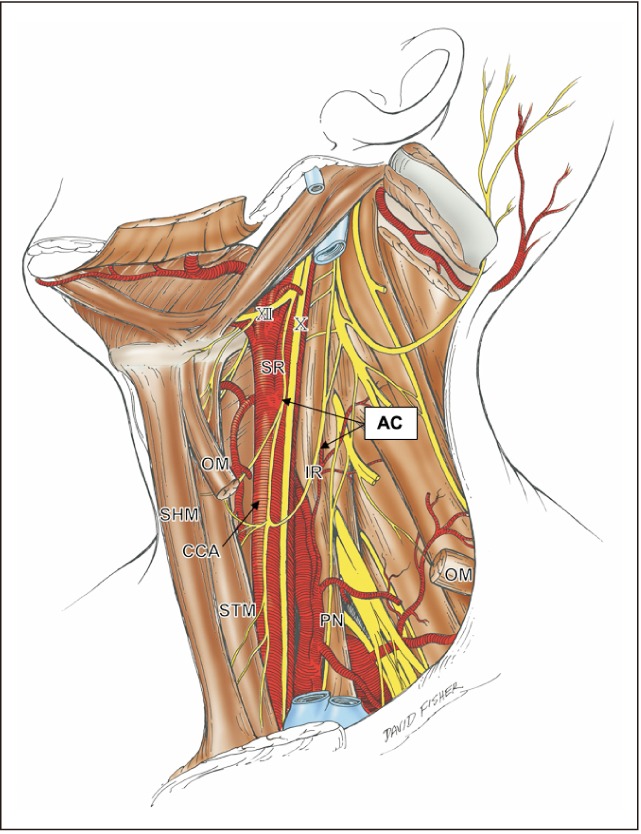
Fig. 1. Schematic drawing of anatomical structures of the ansa cervicalis in the left neck. The platysma, sternocleidomastoid, omohyoid muscle, all veins and the submandibular gland were already dissected. AC, ansa cervicalis; CCA, common carotid artery; IR, inferior root; OM, omohyoid muscle; PN, phrenic nerve; SHM, sternohyoid muscle; SR, superior root; STM, sternothyroid muscle; X, vagus nerve; XII, hypoglossal nerve.

