. 2019 Aug 26;52(3):278–285. doi: 10.5115/acb.19.020

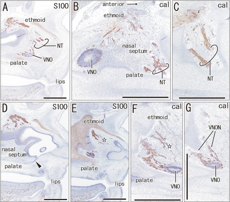
Fig. 1. The terminal nerve and nerves to the vomeronasal organ (VNON) are closely located in a specimen of crown rump length 45 mm (9 weeks). Sagittal sections. Immunohistochemistry of S100 protein (S100) (A, D, E) or calretinin (cal) (B, C, F, G). Panels A and D display the lateral planes in the figure, while panels B, C, F, and G display the medial planes. Thus, the upper or lower raw of panels exhibits a different side from the lateral to the medial, respectively. Intervals between panels are 0.2 mm (A–B), 0.1 mm (B–C), 0.2 mm (D–E), and 0.1 mm (E–F, F–G). On one side (panels A–C), the proximal course of the VNON was not cut, and we did not identify the distal course of the terminal nerve (NT) on the other side (panels D–G). Open stars in panels E and F indicate the most likely candidate of the terminal nerve. Arrowhead in panel D indicates an on-going fusion between the nasal septum and palate. In the specimen, the terminal nerve and VNON appeared to be closely located in the nasal septum. VNO, vomeronasal organ. Scale bars=1 mm (A–G).

Fig. 1