. 2019 Aug 26;52(3):278–285. doi: 10.5115/acb.19.020

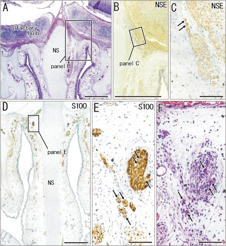
Fig. 5. Ganglion cells on the lateral surface of the crista galli of the ethmoid in a specimen of crown rump length 160 mm (18 weeks). Frontal sections. Panel A is 2.0 mm anterior to panel D. Panel B (or C) is a higher magnification view of a square in panel A (or B). Panel E is a higher magnification view of a square in panel D. Panel F is a section near panel E. Hematoxylin and eosin staining (A, F) or immunohistochemistry of neuron specific enolase (NSE) (B, C) and S100 protein (S100) (D, E). Ganglion cells on the crista galli are, 15–20 µm in diameter, positive for NSE (arrows in panels C), negative for S100 (arrows in panel E), and highly eosinophilic (arrows in panel F). Scale bars=1 mm (A, B, D), 0.1 mm (C, E, F). NS, nasal septum.

Fig. 5