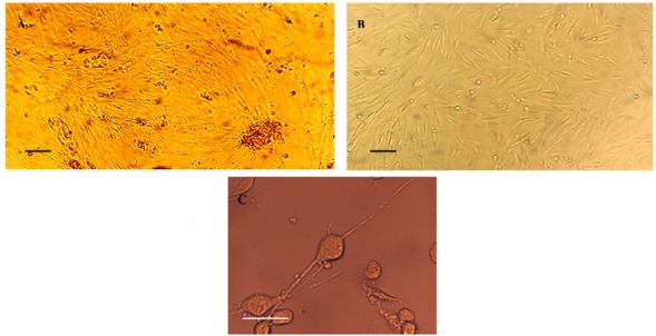
Figure 1.

Phase-contrast microscopic illustration of the cells extracted from Wharton’s Jelly. (A) Attachment and proliferation of spindle-shaped cells occurs with an inside-to-outside direction after culturing the isolated tissue of Wharton’s Jelly. (B) The extracted WJMSCs in the second passage. (C) An illustrative figure of neural-like cells derived from WJMSCs at about 6 hours after initiation with neural induction medium. Figures of treated WJMSCs with VPA are not illustrated. (Scale bars: 20 µm).
