
Figure 2.
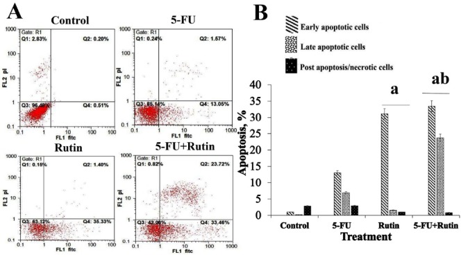
Effect of 5-FU and rutin on the apoptosis of PC3 cells. (A) Dot-plots from flow cytometric illustrating apoptotic status in PC3 cells. (B) Total percentage of apoptosis in PC3 cells treated with the indicated concentrations of 5-FU (0.75 µM) and rutin (700 µM) or a combination of 5-FU with rutin (0.75 µM and 700 µM respectively) for 48 h. All data were expressed as the mean ± standard deviation. aP < 0.05 vs. control cells. bP < 0.05 vs. 5-FU treated cells.