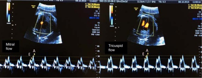
Fig. 5. Spectral Doppler waveforms across the atrioventricular valves (mitral and tricuspid).
Note that the Doppler waveforms are quantified by the E/A ratio, representing fetal diastolic function. The E wave represents passive ventricular filling, while the A wave represents active ventricular filling associated with atrial contraction.

