Figure 3.
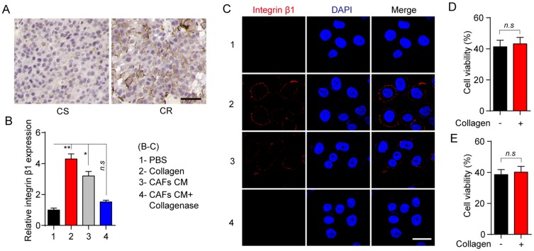
Collagen induces the microtubule-directed chemotherapeutic drugs resistance through the integrin β1. (A) The immunohistochemistry of integrin β1 in breast tumor tissues from CR and CS patients. The scale bar is 50 μm. (B) Relative expression of integrin β1 in MCF-7 pre-treated with collagen (0.5 mg/mL, 12 hrs), CAFs cultured medium (12 hrs) and CAFs cultured medium with collagenase (0.05 mg/mL, 12 hrs). (C) The immunofluorescence of integrin β1 in MCF-7 pre-treated with collagen (0.5 mg/mL, 12 hrs), CAFs cultured medium (12 hrs) and CAFs cultured medium with collagenase (0.05 mg/mL, 12 hrs). The scale bar is 15 μm. (D, E) The cell viability of integrin β1 knockout MCF-7 treated with PBS or collagen (0.5 mg/mL, 12 hrs) to 2 μM PTX (D) and 1nM VCR (E) for 24 hrs. The data were presented as the mean ± SEM from three independent experiments. *P<0.05 and **P<0.01.
Abbreviations: n.s, not statistically significant; CAF, cancer associated fibroblasts; PTX, paclitaxel.

