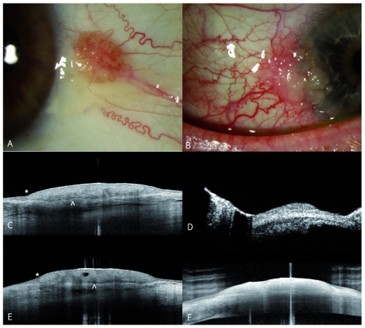
Figure 4. Ocular surface squamous neoplasia (OSSN).
Written informed consent was obtained from the patient in Figure 4, for the use and publication of these images. A) External photograph of left eye showing OSSN with large feeder vessel and surrounding pinguecula temporal to limbus. C) Horizontal and E) vertical anterior segment (AS) OCT scans demonstrating abrupt transition to elevated epithelial lesion (asterisk) and clear plane of separation from underlying tissue (white arrow). B) External photograph of a right eye with OSSN lesion extending from temporal limbus onto cornea. AS-OCT of the lesion ( F) demonstrates significantly higher resolution than ultrasound biomicroscopy of the same lesion ( D).

